

- IJBMR - Aim and Scope
- IJBMR - Open Access
- IJBMR- Journal Details
- Why Publish in IJBMR?
- IJBMR Indexing
- Licenses
- Categories
- IJBMR Performance
- Publication Ethics
- Generative AI Statement
- Revenue Sources
- Advertising Principle
- Direct Marketing Principle
- Archiving Principle
- Privacy Statement
- Instruction for Author
- Author Registration
- Submission
- Copyright Document
- Copyright and Licensing
- IJBMR - Model Covering Letter
- Publication Charges
- Peer - Review Policy
- Conflict of Interest
- Plagiarism
- Journal Policy - Publication Ethics and Malpractice Statement
International Journal of Biological and Medical Research (IJBMR)
²Department of Medical Laboratory Science, Plateau State University State, Nigeria
Department of Medical Laboratory Science,
University of Jos,
Plateau State, Nigeria
ORCID http://orcid/0000-0002-7631-8782 E-mail: solomonmatthias85@gmail.com
Oxidative stress results when reactive oxygen species (ROS) are excessively produced and antioxidants available are not able to counteract this imbalance [1]. Increased levels of free radicals, an imbalance in the body's synthesis and removal of active species, and a weakening of the antioxidant defence system are the outcomes of oxidative stress. Elevated oxidative stress can lead to cellular structural damage and even tissue destruction [2]. Although ionising radiation therapy has many therapeutic advantages for people, it is also associated with the risk of its detrimental effects. By directly depositing energy into macromolecules or radiolyzing water, ionising radiation causes cellular damage to biological systems. This leads to an overabundance of reactive oxygen species (ROS), such as superoxide radicals (O2·−), hydroperoxyl radicals (HO2·), hydroxyl radicals (OH·), and many more examples. These ROS in turn, react with biomolecules and cause oxidative damage to them. Responses to ionising radiation exposure vary greatly and are influenced by factors such as tissue susceptibility, period of exposure, interval between exposures, and absorbed dose [3].
Oxidative stress results in lipid peroxidation, protein changes and DNA damage, sperm death and alterations in blood cells [1]. Oxidative stress is shown by changes in antioxidant status and changed activity of cellular enzymes, such as superoxide dismutase (SOD), catalase (CAT), glutathione peroxidase (GPx) and glutathione which is a non-enzymatic antioxidant [4].Oxidative stress has an impact on a wide range of cellular processes such as metabolism, signalling pathways, regulatory pathways for gene expression, cell proliferation and programmed cell death. Increased levels of free radicals affect the structure and function of the primary biological molecules in the body, such as proteins, lipids, and nucleic acids, and eventually cause damage to tissue. Oxidative stress may affect the functions of blood cells, the coagulation system, and the lipid profile, resulting in acute and chronic infections, anaemia, and hypercoagulability. Complete blood count reflects the cellular immune response[2].
The spleen has a high metabolic rate because of leukocyte activity, making it susceptible to oxidative stress and injury.It has been proven that the interaction of ionising radiation with the biological system leads to increased intracellular ROS, resulting in mutations and chromosomal abnormalities in bone marrow and spleen of mice [5]. There have also been reports of histomorphological changes in the spleen of rats exposed to gamma radiation. In mice exposed to X-rays, there have been reports of a decrease in splenic lymphocytes. Epidemiological studies have shown that ionising radiation can cause a persistent inflammatory state, a dose-dependent immune response impairment, and a dysregulation of cytokine production [6]. According to a study by Dainiaket al.[2], ionising radiation may cause a significant loss of blood cells, which could damage wound healing, inhibit immunological response, and raise the risk of infection. Ionising radiation not only suppresses the immune system but also triggers the release of proinflammatory cytokines such as interleukin-6 (IL-6) and tumour necrosis factor-α (TNF-α), which can result in autoimmune conditions. This highlights the significance of the spleen in this study.
The chaya leaf is a medicinal plant, and because they have a wide range of pharmacological characteristics and chemical structures, medicinal plants have been shown to be a valuable resource in the quest for novel pharmaceuticals.Phenolic compounds, which are a major component of Chaya leaf, can work at several molecular levels and cover a broad spectrum of metabolic activities. These are found in medicinal plants and perform a multitude of physiological functions in the body, including platelet aggregation inhibition, antioxidant, and anti-inflammatory effects. Therefore, there are health benefits linked to the consumption of phenolic substances [7].
A total of 18 healthy weaned albino rats of two weeks old weighing 70-75g were used for this study. The rats were allowed to acclimatize to the environment for one week and maintained under the controlled conditions of temperature (23 ± 2°C), humidity (50 ± 5%), and 12hr of light and dark cycle, respectively. Animals were divided into four groups (A, B, C, D). Group A and B serving as the control (positive and negative control) consisting of 3 rats each while group C and D as the experimental group consisting of 6 rats each, which were exposed to irradiation at low dose, and high dose (75KVP 1MAS, and 115KVP3MAS) respectively at the source target of 70 cm. The average weights and water consumption were recorded. Sawdust was used for bedding and changed regularly during the experiment to ensure air circulation and the animals' free movement.
X-ray irradiation was carried out at the Department of Radiology, National Veterinary Research Institute, Vom, Jos Plateau State, Nigeria, using high-voltage tank model number A6544- 02. Ethical approval and considerations were fully complied with.
At the end of the experiment, all animals were painlessly sacrificed under chloroform anesthesia and subjected to standard necropsy procedures. The appearance and macroscopic changes of in-situ organs, mucosal and serous membranes were observed.
Oxidative stress was established using blood collected from the retro-orbital plexus. The biomarkers used include reduced Glutathione (GSH) activity was estimated in blood serum [8]; Catalase activity was determined using the method of Claiborne [9]; SOD activity was determined in blood serum by the method of Misra and Fridovich[10]; Lipid peroxidation was determined by measuring the levels of Malondialdehyde produced during lipid peroxidation according to the method described by Varshney and Kale [11]. The spleen was excised, fixed in 10% formal saline and processed into paraffin wax. Thin sections of 5μm were cut and stained by the Haematoxylin and Eosin method. Stained sections were examined microscopically [12]. Total leucocyte count, differential leukocyte count, and total platelet count were carried out using the haematologyanalyser. The aqueous extract of Cnidoscolus chayamansa leaves was prepared using Cold Maceration method [13].
PHYSICAL OBSERVATION
Table 1: General Observations Of The Animal During The Period Of The Experiment.
|
Group |
Treatment |
Physical and Behavioural Observations |
|
|
A |
Negative control |
Rats were active, had smooth fur, normal eye color, and Normal urine production. |
|
|
B |
Positive control |
Rats had smooth fur, sluggish for about hour after administration of C. chaya extract, loose faecal droppings. |
|
|
C |
Low dose X-ray Irradiation |
After exposure
After treatment
|
Rats were restless during the first irradiation when compared to the control group, and this restlessness increased significantly during the second irradiation. Their eyes, urine, and feces were all normal.
Faecal droppings were loose, rats were weak for about 60 minutes following the administration of C. chaya extract, smooth fur, normal eye color, and normal urine output. |
|
D |
High dose X-ray Irradiation |
After exposure
After treatment |
Rats were restless during first irradiation when compared to the control group, and this restlessness significantly increased during the second radiation. Their eyes and urine were normal. They produced more feces and urine. Faecal droppings was loose, rats were weak for about an hour after the administration of C.chayamansa extract, smooth fur appearance, normal eye color and normal urine. |
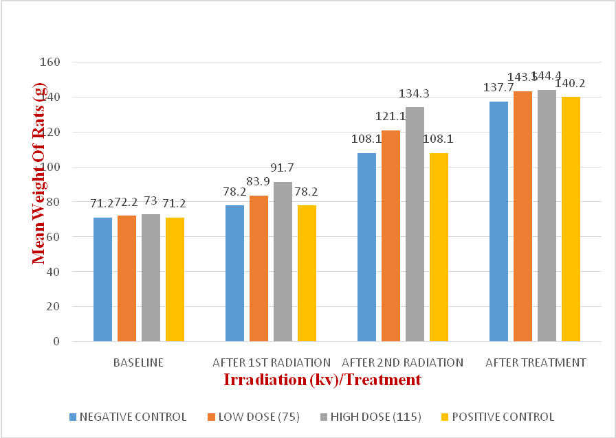
Fig 4.1 Bar chart showing the mean weight of rats (g) throughout the experiment across various irradiation groups.
This demonstrates that all the animals are roughly the same weight at baseline, but following the first irradiation, the animal receiving low-dose radiation experienced a significant increase in weight relative to the control group. This effect was also seen following the second irradiation, but following treatment, the weight difference appears to be decreasing. When compared to the control group and the group receiving low-dose irradiation, the animal receiving high-dose irradiation also showed an increase in body weight; this was also seen after the second irradiation; however, following treatment, the weight difference appears to be decreasing when compared to the control group.
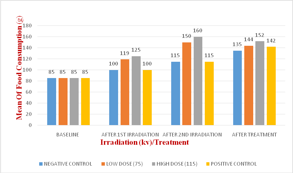
Fig 4.2 Bar chart showing the mean food consumption of rats (g) throughout the experiment across various radiation groups.
All groups' baseline food consumption was normal, and the control group's food intake stayed normal for the period. As a result of the radiation remission, group C saw an increase in food consumption, which is consistent with group D's observation that food consumption increases as radiation dosage increases. However, food consumption in groups C and D dropped throughout the treatment following administration of C. chayamansa leaf extract.
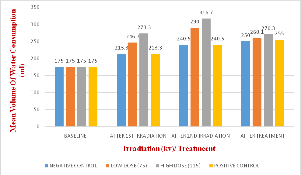
Fig 4.3 Bar chart showing the mean volume of water consumption of rats (ml) throughout the experiment across various radiation groups.
For the duration of the trial, the control group's water consumption stayed constant. As a result of the radiation remission, group C saw an increase in food consumption, which is consistent with group D's observation that food consumption increases as radiation dosage increases. However, food consumption in groups C and D dropped throughout the treatment following administration of C. chayamansa leaf extract.
-
- BIOMARKERS OF OXIDATIVE STRESS
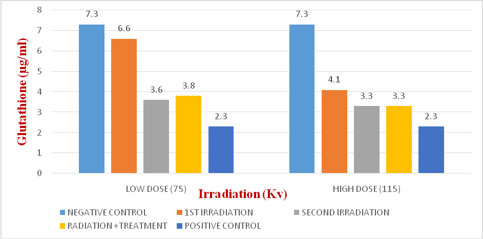
Fig 4.4 Bar chart showing the glutathione level of the animal before radiation, after first radiation, after second radiation and after treatment for both the high and low dose radiation groups
It is observed in figure above that the level of glutathione (GSH), an important antioxidant remained the same throughout the experiment in the control group (A), but there is a decline in the level of glutathione after whole body irradiation in both the animal that received high dose irradiation (D) and low dose irradiation (C), which became slightly increased after the animals were treated with C. chayamansa leaf extract. It is also observed that animals in group B which are the positive control, also had a decrease in GSH level after the treatment with the leaf extract.
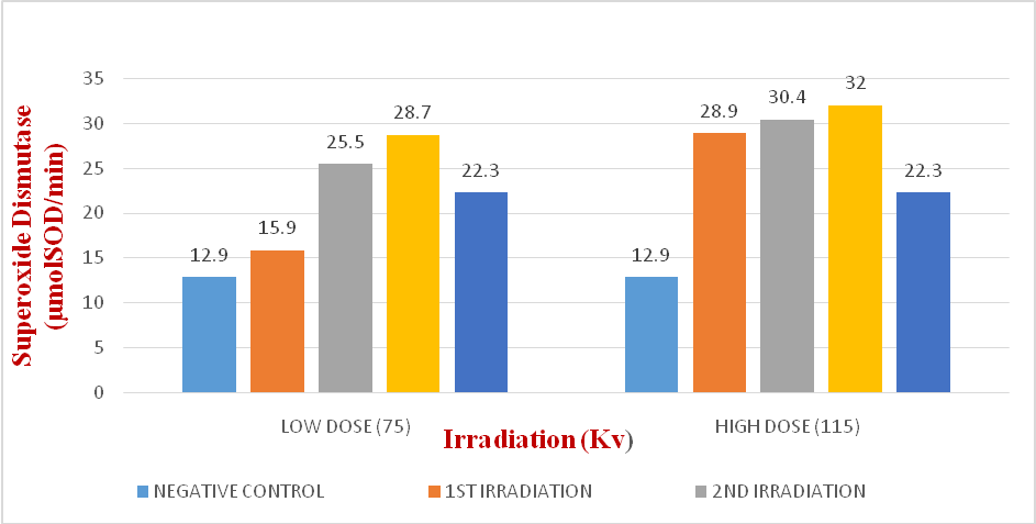
Fig 4.5 Bar chart showing the superoxide dismutase level of the animal before radiation, after first radiation, after second radiation and after treatment for both the high and low dose radiation groups.
The level of superoxide dismutase in the control group (A) was found to remain constant throughout the experiment. However, after whole body irradiation, both the animals that received low dose (C) and high dose (D) radiation showed an increase in superoxide dismutase levels; this increase was further observed after the animals were given C. chayamansa leaf extract. It was also noted that, in comparison to the negative control, the animal in group (B), which functioned as the positive control and was given only the leaf extract, had a higher level of superoxide dismutase.
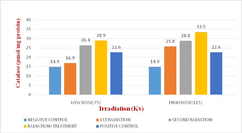
Fig 4.6 Bar chart showing the catalase levels of the animal before radiation, after first radiation, after second radiation and after treatment for both the high and low dose radiation groups
The level of catalase in the control group (A) was found to stay constant during the experiment, but after whole body irradiation, both the animal receiving high dose irradiation (D) and the animal receiving low dose irradiation (C) showed an increase in catalase levels. This increase was noted after the treatment was administered to both irradiation groups. In addition, it was noted that, in comparison to the negative control, the animals in group B, the positive control, had higher levels of catalase following the leaf extract treatment.
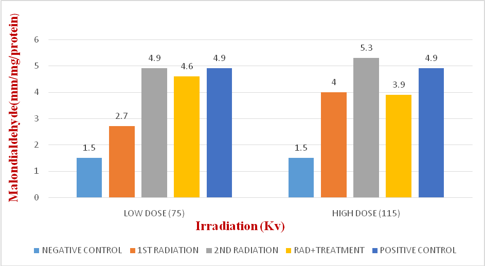
Fig 4.7 Bar chart showing the malondialdehyde level of the animal before radiation, after first radiation, after second radiation and after treatment for both the high and low dose radiation groups.
The level of malondialdehyde in the control group (A) was found to stay constant during the experiment, but after whole body irradiation, the levels of malondialdehyde increased in the animals that received both low dose (C) and high dose (D) radiation. After treatment with C. chayamansa leaf extract, a decrease in malondialdehyde levels was seen in both irradiation groups. Additionally, it was noted that, in comparison to the negative control, the animals in group B, the positive control, likewise showed an increase in malondialdehyde levels following the leaf extract treatment.
4.4 HAEMATOLOGY RESULTS
|
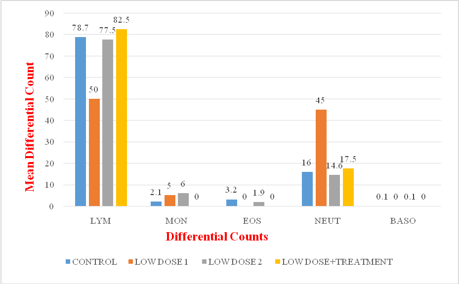
Fig 4.8 Bar chart showing differential count of animals that received low dose irradiation |
Key:
LYM- Lymphocyte
MON- Monocytes
EOS- Eosinophil
NEUT- Neutrophil
BASO- Basophil
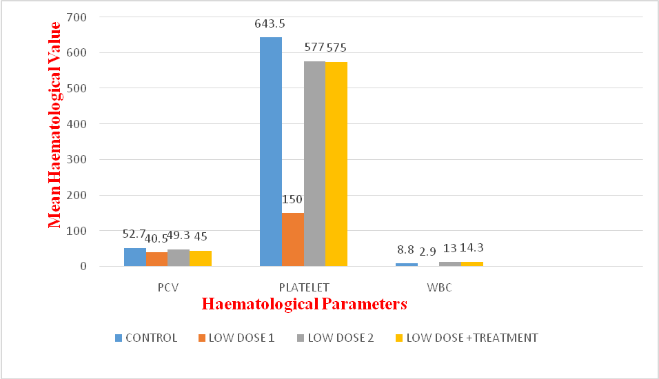
Fig 4.9 Bar chart showing haematological parameters of animals that received low dose irradiation.
Key:
PCV- Parked Cell Volume
WBC- White blood cell
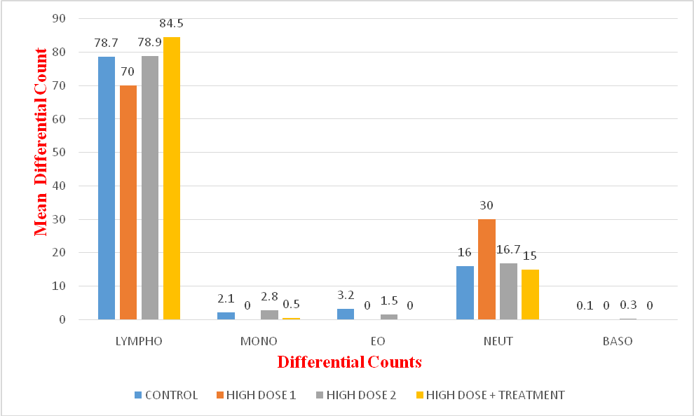
Fig 4.10 Bar chart showing differential count of animals that received high dose irradiation.
Key:
LYM- Lymphocyte
MON- Monocytes
EOS- Eosinophil
NEUT- Neutrophil
BASO- Basophil
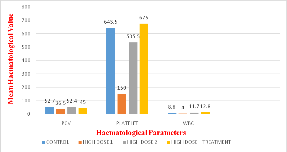
Fig 4.11Bar chart showing haematological parameters of animals that received high dose irradiation.
Key:
PCV- Parked Cell Volume
WBC- White blood cell
4.3 HISTOLOGICAL RESULT
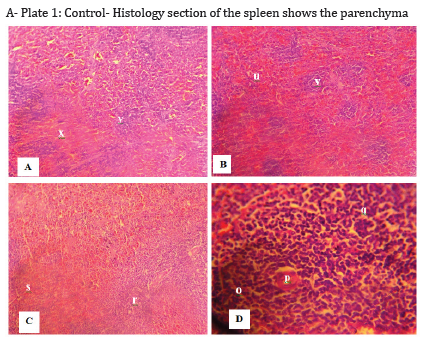
A- Plate 1: Control- Histology section of the spleen shows the parenchyma consists of red pulp (x), with small white nodules of lymphatic tissues scattered throughout called the white pulp (y). H&E, X100
B- Plate 2: Low dose after first irradiation- Histologic section of the spleen shows the parenchyma consisting of red pulp (u), with more frequently occurring small white (v) nodules of lymphatic tissues. H&E X100
C- Plate 3: Low dose after second irradiation- Histologic section of the spleen shows dispersed presence of moderately sized white pulp (r) in the setting of red pulp (s). H&E. x100
D- Plate 4: Low dose after treatment with CnidoscolusChayamansa - Histologic section of spleen shows a prominent white pulp (o) with the characteristics central artery(p) and cluster of white cells (q). This section does not show the relative proportion of white and red pulp. X400 H&E
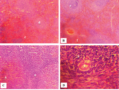
A- Plate 5: Control- Histologic section of the spleen shows the parenchyma consisting of red pulp (c), with more frequently occurring small white nodules (d) of lymphatic tissues. (H&E X100)
B- Plate 6: High dose after first irradiation- Histologic section of the spleen showing clusters of white pulp (e) amid red pulp (f). (H&E X100)
C- Plate 7: High dose, after second irradiation- Histologic section of the spleen shows a spread out of white pulp (g) leaving an ill-defined red pulp (h). (H&E X100)
D- Plate 8: High dose after treatment with Cnidoscoluschayamansa - Histological section of spleen shows the white pulp (k) with a wide margin of lymphocytes, surrounding the central artery (l). There is a small portion of the red pulp that unremarkable. (H&E X400).
4.4 HAEMATOLOGICAL RESULTS
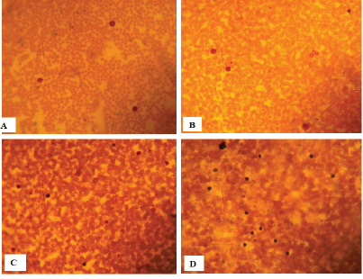
Plate A: No irradiation exposure. Microcytic polychromatic cells, neutrophil left shift present with adequate number of platelets.
Plate B: First exposure at high dose. Polychromatic, microcytic, neutrophil left shit present with adequate number of platelets.
Plate C: Second exposure at high dose. erythrocytosis, lymphocytosis, anisocytosis, microcytosis, reactive lymphocyte, polychromatic cells.
Plate D; Post treatment with Cnidoscolus chayamansa. Polychromatic cells and microcytic cells. Elevated platelet number.
GENERAL OBSERVATIONS
The rats' physical and behavioural responses generally indicated the toxic and lethal consequences of whole-body irradiation. Table 4.1 shows the general behaviour of the rats as well as their activities during the experiment. At baseline, all rats were active, alert, and exhibited normal eye color, smooth fur, and regular fecal droppings. The negative control group (A) showed no abnormal behavior, with normal fur, urine output, and activity. In contrast, the irradiation and treatment groups displayed significant changes in alertness, fecal texture, and fur appearance. These effects are likely due to reactive oxygen species from irradiation [14]. The treatment group's recovery signs may be linked to bioactive compounds and the antioxidant flavonoids in C. chayamansa. Research indicates that the plant is rich in flavonoids and other phytochemicals known for their antioxidant properties [15].
After treatment with C. chayamansa leaf extract, rats in group C (low-dose irradiation) showed normal eye colour, urine output, and faecal production. However, alertness was higher during the first irradiation than the second. Faeces became loose, and rats exhibited weakness for about an hour post-treatment, though fur remained smooth, and eye colour and urine output stayed normal.
Animals in group D (high-dose irradiation) were notably more restless and active during the first irradiation compared to the control group, with a marked increase during the second. They exhibited normal eye colour, normal urine, and increased faecal and urine output. Following C. chayamansa extract treatment, rats exhibited loose faeces, transient weakness (~1 hour), smooth fur, normal eye colour, and urine output. According to Giday et al. [16], the laxative effect may be due to phytochemicals that increase intestinal water content or stimulate motility. The transient weakness is likely a result of the body's metabolic response to the plant's bioactive compounds, possibly involving a temporary energy depletion or central nervous system modulation. Also, the typical markers of systemic health, smooth fur, normal eye colour, and typical urine output, suggest that while C. chayamansa causes some immediate effects, but it does not result in lasting harm at the doses used. This observation aligns with other studies on medicinal plants that are generally deemed safe but may cause temporary physiological reactions as the body adjusts to the intake of new compounds [17].
Figure 4.1 shows an increase in mean body weight during the irradiation period. Groups C and D started with similar weights, but both gained significantly after the first irradiation compared to controls, with Group D gaining more than A, B, and C. This supports Balan et al. [18], who suggested that oxidative stress may trigger fat storage as a compensatory response.
After treatment, the animals lost weight at a rate similar to the control group, likely due to reduced appetite and increased metabolic demand during recovery from irradiation damage [14]. Group B (the positive control) showed slight weight gain compared to the negative control. Although the plant extract may provide antioxidant benefits, it does not directly counteract irradiation-induced weight loss, aligning with studies indicating that such plants primarily reduce oxidative stress and inflammation rather than maintain weight [19]. Figure 4.2 shows normal feeding at baseline. After the first irradiation, animals receiving low doses displayed increased appetite compared to controls, a trend that continued after the second exposure. Animals exposed to high-dose irradiation also showed greater feeding than both the control and low-dose groups, even after second irradiation. These findings align with Balan et al. [18], who reported that oxidative stress disrupts the hormones leptin and ghrelin, which control appetite and satiation. This imbalance may result in less energy being expended and more food being consumed, which would add to the weight gain. The increase in food intake following both low and high-dose irradiation may represent an adaptive response to increased metabolic demands for tissue repair and recovery from irradiation-induced damage [20]. In contrast, after therapy, animals exhibited reduced appetite compared to controls. This suggests that the antioxidant properties of C. chayamansa may have mitigated oxidative stress and contributed to the restoration of normal appetite-regulating mechanisms. By reducing oxidative damage, the extract may help rebalance leptin and ghrelin activity, leading to normalisation of feeding behaviour[19].
Water intake remained stable in the control group throughout the experiment (Figure 4.3). Following irradiation, groups C and D showed increased water intake, with higher intake corresponding to higher irradiation doses. However, after treatment with C. chayamansa leaf extract, water intake in both groups gradually decreased over the treatment period (Figure 4.3).
BIOMARKERS OF OXIDATIVE STRESS
Figure 4.4 shows that glutathione (GSH) levels remained stable in the control group (A) throughout the experiment. However, GSH levels declined following whole-body irradiation in both the low-dose (C) and high-dose (D) groups. Glutathione levels decreased progressively with increasing doses of X-ray irradiation, with significantly lower values in the high-dose group compared to the low-dose group. A further decline was observed in both groups after the second irradiation. This supports Sener et al. [21], who reported that GSH plays a critical role in protecting against oxidative injury and is often depleted under oxidative stress, allowing greater free radical-induced damage. However, this contrasts with Gunes et al. [22], who suggested that antioxidant enzymes generally increase their activity under oxidative stress. Group B (positive control) also showed a decrease in GSH levels after treatment with the leaf extract, suggesting that irradiation reduced antioxidant levels and the extract, despite its antioxidant properties, may have further influenced this decline.
As shown in Figure 4.5, rats in the control group (A) maintained normal superoxide dismutase (SOD) levels throughout the experiment. In contrast, SOD levels increased progressively in both low- and high-dose irradiated groups, with higher doses resulting in greater increases, that is the higher the dose of irradiation the more the level of superoxide dismutase. Superoxide dismutase (SOD), a key antioxidant enzyme, increases in response to oxidative stress, confirming the presence of oxidative stress [22,23]. Group B (positive control) also showed elevated SOD levels after treatment with the leaf extract, suggesting that irradiation-induced oxidative stress triggered antioxidant activity, and the extract may have acted synergistically with the body’s defense mechanisms to enhance free radical scavenging.
Figure 4.6 shows that catalase levels remained stable in the control group (A) throughout the experiment. However, catalase levels increased following whole-body irradiation in both the low-dose (C) and high-dose (D) groups. Catalase levels increased progressively with rising doses of X-ray irradiation, as animals in the high-dose group exhibited higher levels than those in the low-dose group. Both groups also showed further elevation after the second exposure. This supports the findings of Gunes et al. [22], which emphasized that oxidative stress such as that caused by ionizing irradiation induces upregulation of antioxidant enzymes like catalase as part of the body’s defense mechanism against reactive oxygen species (ROS). Similarly, an increase in catalase activity in Group B (positive control) following treatment with the leaf extract suggests a synergistic effect, where the extract’s antioxidant compounds may have enhanced the body’s endogenous antioxidant response to counteract oxidative damage.
The level of malondialdehyde (MDA) in the control group (A) remained stable throughout the experiment. However, MDA levels increased significantly following whole-body irradiation in both the low-dose (C) and high-dose (D) groups, with a markedly higher concentration observed in the high-dose group. This indicates that irradiation dose plays a critical role in the extent of oxidative stress. As an established marker of oxidative damage, elevated MDA levels reflect increased production of reactive oxygen species [24]. Post-treatment with C. chayamansa leaf extract, a reduction in MDA levels was noted in both irradiated groups. In contrast, Group B (positive control) showed a slight increase in MDA levels after treatment compared to the negative control.
HISTOLOGICAL OBSERVATION
1: The Spleen
The splenic histologic section of the control group after first irradiation as presented in Plate1 showed normal splenic structure characterized by the parenchyma consisting of red pulp, with small white nodules of lymphatic tissues called the white pulp scattered throughout called the white pulp.
The histology of the spleen after first irradiation of low dose of 75KV/1mAs as represented in Plate 2, showed the parenchyma consisting of red pulp, with more frequently occurring small white nodules of lymphatic tissues. On second exposure in Plate3, it showed dispersed presence of moderately sized white pulp in the setting of red pulp. This agrees with the work done by Balaet al. [3], who established that when ionizing radiation interacts with the biological system, it increases intracellular Reactive Oxygen Species (ROS), which causes chromosomal alterations and mutations in the mice's spleen and bone marrow.
The histology of the spleen after first irradiation of high dose of 11kv/3mAs (Plate 6) showed clusters of white pulp in the midst of red pulp. And on second exposure (plate 7) it showed a spread out of white pulp leaving an ill-defined red pulp. While this agrees with the study by Balaet al. [3], this disagrees with Jin H et al. [25] observations that histopathological analysis of splenic tissue of mice exposed to X-ray irradiation exhibited a reduction in the amount of white pulp and an increased red pulp area.
On treatment with C. chayamansaevery day for 10 days, the histology of the spleen exposed to low dose irradiation showed a prominent white pulp with the characteristic central artery and cluster of white cells (plate 4a and 4b) and an ill-defined red pulp due to the widespread presence of lymphocytes. While the histology of the spleen exposed to high-dose irradiation showed the white pulp with a wide margin of lymphocytes, surrounding the central artery with an unremarkable small portion of the red pulp (plates 8a and 8b). This shows remarkable features of recovery from the effects of oxidative stress. This is supported by a study carried out by Mbahet al., [26], that flavonoids present in C. chayamansa leaf extract can neutralize free radicals. These flavonoid substances aid in counteracting free radicals, leading to a decrease in oxidative stress and the prevention of chronic illnesses.
HAEMATOLOGICAL PARAMETERS
After whole-body gamma irradiation, injury to animal tissues was well reflected in the peripheral blood. Lymphocytes are highly radiosensitive, and their depletion is commonly associated with a weakened immune response. In this study, a reduction in lymphocytes was observed after the second low-dose irradiation, consistent with findings by Jin et al. [25], who reported decreased splenic lymphocytes following X-ray exposure in mice. Interestingly, lymphocyte levels increased under high-dose exposure and further increased in both irradiation groups after treatment with C. chayamansa extract. This suggests a potential immunomodulatory effect of the extract in restoring lymphocyte levels post-irradiation. The administration of C. chayamansa appears to promote lymphocyte recovery or proliferation, likely due to its antioxidant properties. By mitigating irradiation-induced oxidative stress, these antioxidants may support lymphocyte regeneration. This observation aligns with previous studies reporting that plant extracts rich in antioxidants can enhance immune function and promote lymphocyte survival and proliferation following irradiation [19,22].
White blood cell (WBC) counts increased across both irradiation doses, consistent with findings by Bala et al. [3] who reported elevated WBC levels in X-ray–exposed mice compared to controls. This increase persisted following treatment with C. chayamansa, suggesting the extract may possess immunomodulatory properties. Its antioxidant activity may enhance the body’s capacity to maintain or stimulate leukocyte production in response to irradiation-induced oxidative stress. The elevated WBC count may reflect an activated immune response aimed at repairing and defending against irradiation-induced damage. It could also indicate reduced immunosuppression, a typical effect of ionizing irradiation. Thus, C. chayamansa may support hematopoietic recovery post-irradiation, consistent with its documented antioxidant and protective properties [3,20].
Neutrophil and eosinophil counts decreased after the second low-dose irradiation but increased following the second high-dose exposure. Post-treatment with C. chayamansa, improvements were observed. As noted by Hall and Giaccia[14], such patterns reflect the body's acute inflammatory response to stress or injury. The reduction at low doses likely indicates irradiation-induced suppression of hematopoietic cells, while the increase at high doses suggests a compensatory immune response to extensive tissue damage and inflammation.
Monocyte counts increased after both low- and high-dose irradiation, reflecting a typical inflammatory response to irradiation-induced damage. Ionizing irradiation causes cellular injury and oxidative stress, prompting an immune reaction involving elevated monocytes for tissue repair and defense [26, 27]. However, post-treatment with C. chayamansa, monocyte levels declined in the high-dose group, suggesting a modulatory effect of the extract on inflammation. Monocytes are key players in tissue repair and immune regulation, explaining their proliferation following irradiation exposure. The reduction observed after C. chayamansa treatment in the high-dose group suggests the extract may exert anti-inflammatory effects by alleviating oxidative stress, thereby diminishing the need for an amplified immune response.
Packed Cell Volume (PCV) decreased following both low and high doses of irradiation, indicating haematological alterations due to X-ray exposure. This aligns with Danesh et al. [3], who reported that oxidative stress can cause anaemia, hypercoagulability, infections, and blood cell dysfunctions. Post-treatment with C. chayamansa, PCV levels remained unchanged, suggesting the extract had no immediate corrective effect on irradiation-induced PCV reduction. The observed reduction in PCV suggests potential bone marrow or peripheral blood cell damage, a known effect of ionising irradiation due to disrupted hematopoiesis, which may lead to anaemia or related disorders [28]. The unchanged PCV levels after C. chayamansa treatment may imply that the extract may not mitigate the effects of irradiation on PCV, or that its mechanism of action does not influence this particular parameter.
Platelet counts decreased following both low and high doses of X-ray irradiation, consistent with Bala et al. [3], who reported similar reductions in irradiated animals. This decline suggests irradiation-induced damage to hematopoietic tissues or disruption of normal thrombopoiesis. The increase in platelet counts in the high-dose irradiation group after treatment with C. chayamansa suggests a potential therapeutic effect on irradiation-induced thrombocytopenia. This may be due to enhanced platelet production or reduced destruction, aligning with reports that medicinal plants, including C. chayamansa, can support haematological recovery under stress conditions [29,30].
This study highlights the toxic effects of oxidative stress induced by X-ray irradiation and the therapeutic potential of C. chayamansa. Whole-body irradiation caused notable physiological and behavioural alterations, including changes in platelet counts, body weight, and feeding patterns, which are indicators of irradiation-induced tissue damage. Treatment with C. chayamansa alleviated these effects, as shown by improvements in oxidative stress biomarkers (glutathione, superoxide dismutase, and catalase), histological recovery, and haematological parameters. The plant’s antioxidant properties likely underpin its protective role, suggesting its promise as a radioprotective agent. Further research is warranted to explore its mechanisms of action, its potential clinical applications and identify its most effective dose for treating radiation-induced damage.
The authors thank the Animal House of the University of Josfor providing the ethical clearance for the use of experimental animals to perform the research.
None.
Conflicts of Interest:
The authors declare no conflict of interest.
[1] Agarwal, A., Durairajanayagam, D., Halabi, J., Peng, J., & Vazquez-Levin, M.. Proteomics, oxidative stress and male infertility. Reproductive BioMedicine Online, 2014; 29(1), 32–58. https://doi.org/10.1016/j.rbmo.2014.02.013
[2] Danesh, H., Ziamajidi, N., Mesbah-Namin, S. A., Nafisi, N., &Abbasalipourkabir, R. (2022). Association between Oxidative Stress Parameters and Hematological Indices in Breast Cancer Patients. International Journal of Breast Cancer, 2022, e1459410. https://doi.org/10.1155/2022/1459410
[3] Bala, S., Chugh, N. A., Bansal, S. C., &Koul, A. Aloe veramodulates X-ray induced hematological and splenic tissue damage in mice. Human & Experimental Toxicology, 2019; 38(10), 1195–1211. https://doi.org/10.1177/0960327119860174
[4] Demirci-Çekiç, S., Özkan, G., Avan, A. N., Uzunboy, S., Çapano?lu, E., &Apak, R. (2021). Biomarkers of Oxidative Stress and Antioxidant Defense. Journal of Pharmaceutical and Biomedical Analysis, 209(114477), 114477.https://doi.org/10.1016/j.jpba.2021.114477
[5] ManjuLata Gupta, &Verma, S. Prophylactic strategies to minimize the effect of whole body irradiation on hematopoietic, gastrointestinal and respiratory system leading to morbidity/mortality in animals. Indian Journal of Radiation Research, 2018; 9(1), 4–4. https://doi.org/10.4103/jrcr.jrcr_2_18
[6] Park B, Yee C and Lee KM. The effect of radiation on the immune response to cancer. Int J MolSci 2014; 15: 927–943
[7] Pérez-González, M. Z., Gutiérrez-Rebolledo, G. A., Jiménez-Arellanes, M. A., & Cruz-Sosa, F. The chemical constituents and biological activities of CnidoscoluschayamansaMcVaugh, a Mexican medicinal species, and plant cell cultures for the production of bioactive secondary metabolites. Studies in Natural Products Chemistry, 2021; 317–346. https://doi.org/10.1016/b978-0-12-819485-0.00009-8.
[8] Betteridge, D. J. What is oxidative stress? Metabolism, 2000; 49(2), 3–8. https://doi.org/10.1016/s0026-0495 (00)80077-3
[9] Claiborne, Stephen. “RESPONSE OF HEART RATE VARIABILITY TO PHYSIOLOGICAL STRESS IN HEALTHY COLLEGE STUDENTS.” Master’s thesis, Miami University, 2018. http://rave.ohiolink.edu/etdc/view?acc_num=miami1524782664217834
[10] Misra HP, Fridovich I. The role of superoxide anion in the autoxidation of epinephrine and a simple assay for superoxide dismutase. J Biol Chem. 1972;247(10):3170-5. PMID: 4623845.
[11] R. Varshney and R.K. Kale. Effects of Calmodulin Antagonists on Radiation-induced Lipid Peroxidation in Microsomes. International Journal of Radiation Biology, 1990;58 (5): 733-743
[12] Solomon M. Gamde, C. J. Ugwah-Oguejiofor, A. Garba, G. O. Avwioro, A. Moronkeji, and AbdullahiAbiodunJimoh. “Histologic and Biochemical E1ect of BalaniteaegyptiacaFruit Extract on Alloxan-Induced Diabetes in Wistar Rats, Ethiopian Journal of Health Sciences 2023;33(3): 441–450, https://doi.org/10.4314/ejhs.v33i3.7
[13] Trease, G. and Evans, W. Phytochemicals. In: Pharmacognosy, 15th Edition, Saunders Publishers, London, 2002; 42-393.
[14] Hall, E.J. and Giaccia, A.J. Radiobiology for the Radiologist. 7th Edition, Wolters Kluwer Health/Lippincott Williams & Wilkins, Philadelphia. 2012.
[15] Sathyarengasamy and Ushadevithangaprakasam. Isolation, screening and determination of α-amylase activity from marinestreptomyces species. Int J Pharm PharmSci, 2021;10(4): 122-127. DOI: http://dx.doi.org/10.22159/ijpps.2018v10i4.24447.
[16] Teklehaymanot, T. and Giday, M. Ethnobotanical Study of Wild Edible Plants of Kara and Kwego Semi-Pastoralist People in Lower Omo River Valley, DebubOmo Zone, SNNPR, Ethiopia. Journal of Ethnobiology and Ethnomedicine, 2010; 6, 23. http://dx.doi.org/10.1186/1746-4269-6-23.
[17] Bennett, M. Basic Concepts of Intercultural Communication: Paradigms, Principles, & Practices. Intercultural Press, Boston. 2013.
[18] BijiBalan, Amit S. Dhaulaniya, Munendra Kumar, Mohit Kumar, Prateek Kumar. Aflatoxins in food: Prevalence, health effects, and emerging trends in its mitigation—An updated review. 2024; 2(1):39-71.
[19] ManjeshwarShrinathBaliga, BantwalRaghavendraVittaldasBaliga, Shaun Mathew Kandathil, Harshith P. Bhat d, Praveen Kumar Vayalil. A review of the chemistry and pharmacology of the date fruits (Phoenix dactylifera L.). Food Research International 2011; 44 (2011) 1812–1822.
[20] Choudhary DK, Bhakt P, Kaur R. Erratum for Choudhary et al., “Essential Role for the Phosphatidylinositol 3,5-Bisphosphate Synthesis Complex in Caspofungin Tolerance and Virulence in Candida glabrata”. Antimicrob Agents Chemother. 2019 Oct 22; 63(11):e01794-19. doi: 10.1128/AAC.01794-19.
[21] ?ener, G., Ek?io?lu-Demiralp, E., Çetiner, M., et al. β-Glucan Ameliorates Methotrexate Induced Oxidative Organ Injury via Its Antioxidant and Immunomodulatory Effects. European Journal of Pharmacology, 2006; 542, 170-178. https://doi.org/10.1016/j.ejphar.2006.02.056.
[22] Güne?, S. and Esteves, S. C.. Role of genetics and epigenetics in male infertility. Andrologia, 2020;53(1). https://doi.org/10.1111/and.13586.
[23] Sosa V, Moliné T, Somoza R, Paciucci R, Kondoh H, LLeonart ME. Oxidative stress and cancer: an overview. Ageing Res Rev. 2013:12(1):376-90. doi: 10.1016/j.arr.2012.10.004. Epub 2012 Oct 31. PMID: 23123177.
[24] Xu JY, Zhao L, Chong Y, et al. Protection effect of sanguinarine on whole-body exposure of X radiation in BALB/c mice. Braz J Pharm Sci 2014; 50: 101–106.
[25] Jin, H., Jin, Y. B., Lee, J. W., Kim, J. K., & Lee, Y. S. DIFFERENTIAL EXPRESSION OF RADIATION RESPONSE GENES IN SPLEEN, LUNG, AND LIVER OF RATS FOLLOWING ACUTE OR CHRONIC RADIATION EXPOSURE. Journal of radiation protection and research, 2013; 40(1), 25-35. Musa J., Zakka G. C. and Kachong Y. K. Effect of boiling on the micro and macro mineral composition of Moringa (moringaoleifera) seeds.NIGERIAN SOCIETY FOR ANIMAL PRODUCTION (NSAP) 45TH ANNUAL CONFERENCE - BAUCHI 2020 BOOK OF PROCEEDINGS.
[26] Takai D, Abe A, Miura H, Tanaka IB, Saito M & Komura JI. Adverse effects of chronic low dose-rate gamma-ray exposure ameliorated by environmental enrichment in mice. International Journal of Radiation Research, 2021; 19(3): 543-550.
[27] Ali AbidAbojassim, HaiderSalihJaffat, &AdhraaBaqir Hassan.. EFFECTS OF GAMMA RADIATION ON SOME HEMATOLOGICAL PARAMETERS IN FEMALE RATS. Teoreti?eskaâ I PrikladnaâNauka, 2015; 25(05), 101–109. https://doi.org/10.15863/tas.2015.05.25.19
[28] Mohan, V., Anoop, M., Bhansali, A., Singh, A.K., Makkar, B., Krishnan, D., et al. Role and Significance of Dietary Protein in the Management of Type 2 Diabetes and Its Complications in India: An Expert Opinion. Journal of the Association of Physicians of India, 2023; 71, 36-46.
[29] Loarca-Piña, G., Mendoza, S., Ramos-Gómez, M., & Reynoso, R. Antioxidant, Antimutagenic, and Antidiabetic Activities of Edible Leaves from Cnidoscoluschayamansa Mc. Vaugh. Journal of Food Science, 2010; 75(2), H68–H72. https://doi.org/10.1111/j.1750-3841.2009.01505.xz
[30] Lang, P., H. Eric Xu, Grusdat, M., McIlwain, D. R., Pandyra, A. A., Harris, I. R., NamirShaabani, Honke, N., Sathish Kumar Maney, Lang, E., Pozdeev, V. I., Recher, M., Odermatt, B., Brenner, D. W., Dieter Häussinger, Ohashi, P. S., Hengartner, H., Zinkernagel, R. M., Mak, T. W., & Lang, K. (2013). Reactive oxygen species delay control of lymphocytic choriomeningitis virus. 20(4), 649–658. https://doi.org/10.1038/cdd.2012.167
IJBMR -ISSN: 0976:6685. ©Copyright 2026 This is an open access article under the CC BY-NC-ND license (http://creativecommons.org/licenses/by-nc-nd/4.0/).
Published by BioMedSciDirect Publications
This work is licensed under a Creative Commons Attribution-NonCommercial-NoDerivatives 4.0 International License (CC BY-NC-ND)
